Indications
- Lacerations, foreign body, wound exploration of planar foot
- Calcaneus factures
Contraindications
- Infection overlying injection site
- Allergy to local anesthetic
Equipment
- 5cc of local anesthetic of choice
- 23-25G needle
- Cleansing solution
- Ultrasound with high-frequency linear transducer
- Ultrasound transducer sterile cover
Preparation
Position
Position the patient with slight hip external rotation and knee flexion to expose the medial malleolus
Ultrasound
Landmarks

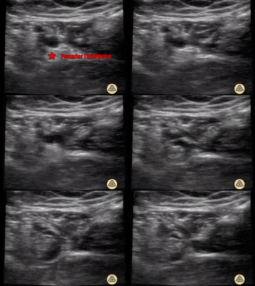
- Place the transducer posterior to the medial malleolus in a transverse orientation
- Identify the tibial artery/vein using doppler flow

- Identify the tibial nerve lateral/posterior to the vascular bundle
Technique
- Introduce needle from posterior aspect
- In-plane needle visualization
- Infiltrate local anesthetic superficial to the tibial nerve (*)

