Indications
- Refractory ventricular arrhythmia
- Non-emergency-department indications described in the source:
- Complex regional pain syndrome
- Sympathetically maintained pain
- Postherpetic neuralgia
- Phantom limb pain
- Post-traumatic stress disorder
- Anxiety
Contraindications
- Infection overlying injection site
- Allergy to local anesthetic
Equipment
- 10 mL of a short-acting local anesthetic, such as 1% lidocaine without epinephrine
- 20–25 G, 5 cm echogenic needle
- Cleansing solution
- Ultrasound with high-frequency linear transducer and color Doppler
- Ultrasound transducer sterile cover
Preparation
Position
Position the patient supine with the head turned to the right to expose the left side of the neck. This may also displace the internal jugular vein and improve the lateral-to-medial needle trajectory.
The source recommends performing the initial block on the left because hypotension has been reported less often after left-sided than right-sided blockade. If there is no improvement after approximately 10 minutes, a contralateral block may be considered based on the clinical context.
Ultrasound
Landmarks
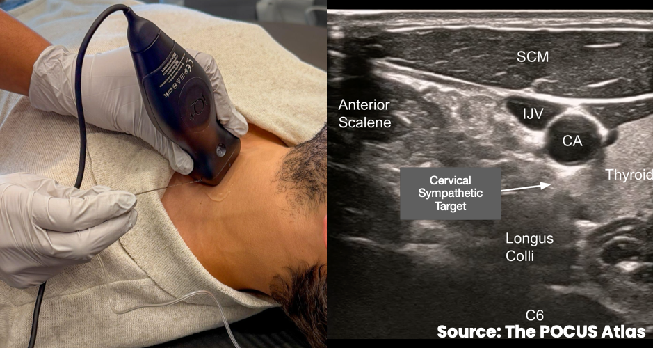
- Place the linear transducer in the transverse plane on the left side of the neck at the level of the cricoid cartilage, approximately C6.
- Identify the sternocleidomastoid muscle, anterior scalene muscle, thyroid, internal jugular vein, carotid artery, and longus colli muscle.
- Use color Doppler before needle advancement and injection to identify the vertebral artery and other vessels in or near the planned trajectory.
- Although the stellate ganglion lies at approximately C7–T1, this ultrasound-guided cervical sympathetic block uses a C6 procedural window to access the prevertebral fascial plane superficial to the longus colli muscle.
Technique
- If chest compressions are in progress, anchor the procedural hand for stability. The needle may be introduced just beneath the skin during compressions and advanced during a pulse check.
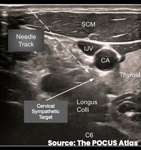
- Advance the needle in-plane from lateral to medial toward the prevertebral fascia along the superficial aspect of the longus colli muscle.
- Maintain direct visualization of the needle and avoid the carotid artery, internal jugular vein, vertebral artery, thyroid, and neural structures.
- Inject 10 mL of short-acting local anesthetic into the target plane while visualizing spread superficial to the longus colli muscle.
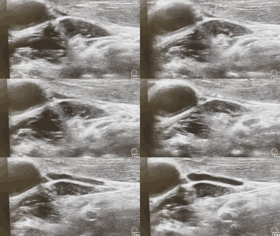
Examples