Indications
- Upper extremity fractures
- Lacerations or abscesses of upper arm
Contraindications
- Infection overlying injection site
- Allergy to local anesthetic
- Vascular injury/injection: There are many large vessels that serve as landmarks so color doppler and negative aspirations are essential
Equipment
- 20-25cc of local anesthetic of choice
- 18-22G needle
- Saline Flush
- Cleansing solution
- Ultrasound with high-frequency linear transducer
- Ultrasound transducer sterile cover
Prepration
Position
- The patient is positioned supine
- Abduction of ipsilateral arm to 90° may aid nerve visualization
Ultrasound
Landmarks
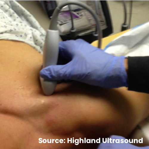
- The probe is placed in a parasagittal orientation between the midpoint of the clavicle and the coracoid process, just inferior to the clavicle.
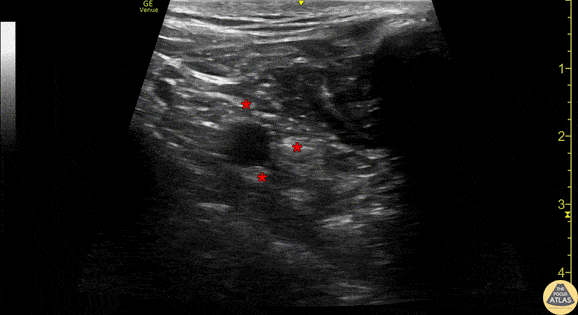
- The brachial plexus cords surround the axillary artery in a variable pattern but cannot always be individually visualized.
Technique
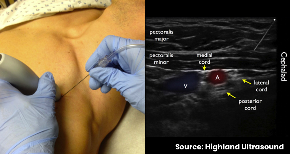
- In-plane needle visualization
- Enter in the parasagittal plane toward the posterior/dorsal aspect of the axillary artery
- Inject local anesthetic in small aliquots just deep to the axillary artery
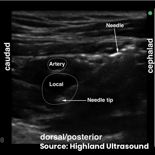
- Correct position can be demonstrated by obtaining the “double bubble” sign as local anesthetic spread in the periplexus space
Example