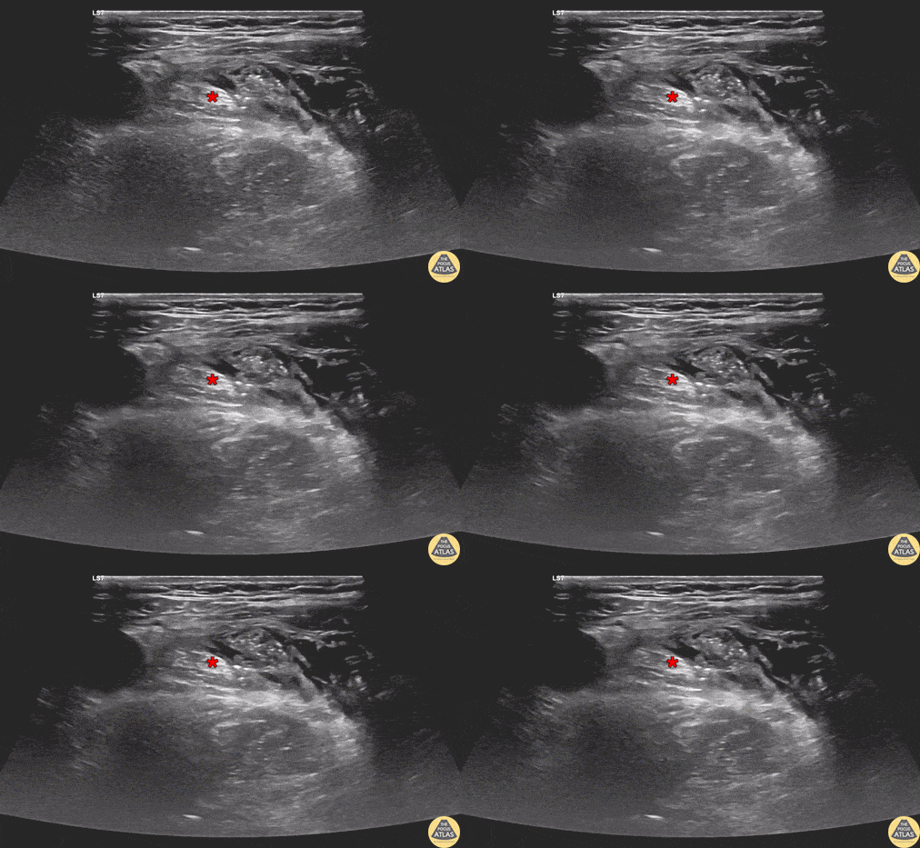
Indications
- Fractures of the femoral head, neck, intertrochanteric/subtrochanteric regions, shaft
- Proximal tibial fractures
- Patella fractures
- Anterior thigh lacerations or abscesses
Contraindications
- Infection overlying injection site
- Allergy to local anesthetic
- Request of consultant
- Concern for compartment syndrome
Equipment
- 15-20cc of local anesthetic of choice
- 22G spinal needle
- Saline Flush
- Cleansing solution
- Ultrasound with high-frequency linear transducer
- Ultrasound transducer sterile cover
Preparation
Position
Position the patient supine
Ultrasound
Landmarks
- Expose the groin to identify the anterior superior iliac spine and inguinal crease
- Place transducer transversly across the femoral region of the upper thigh parallel to the inguinal crease
- The femoral vessels are identified and centered on screen
- Follow the femoral artery proximal to the inguinal ligament and distal to the takeoff of the profunda femoris artery
- The femoral nerve should appear proximal to the bifurcation
Technique
- Find the large nonpulsatile femoral vein as the initial landmark under ultrasound
- Identify femoral nerve (hyperechoic structure lateral to the femoral artery)
- Must also identify fascia lata and deeper fascia iliaca, as these two fascial planes overlie the femoral nerve. If anesthetic is not placed below these two fascial planes, the anesthetic will not reach femoral nerve and the block will fail.
- Under ultrasound guidance, advance needle to the femoral nerve sheath
- Aspirate to ensure not in blood vessel
- Inject 20cc of local anesthetic along nerve sheath
Examples


