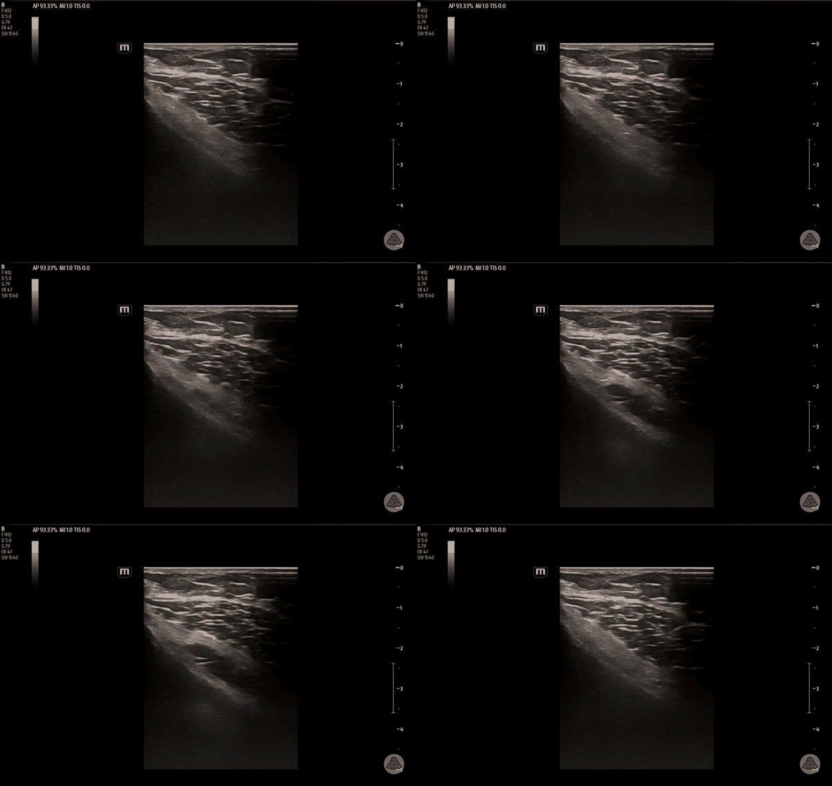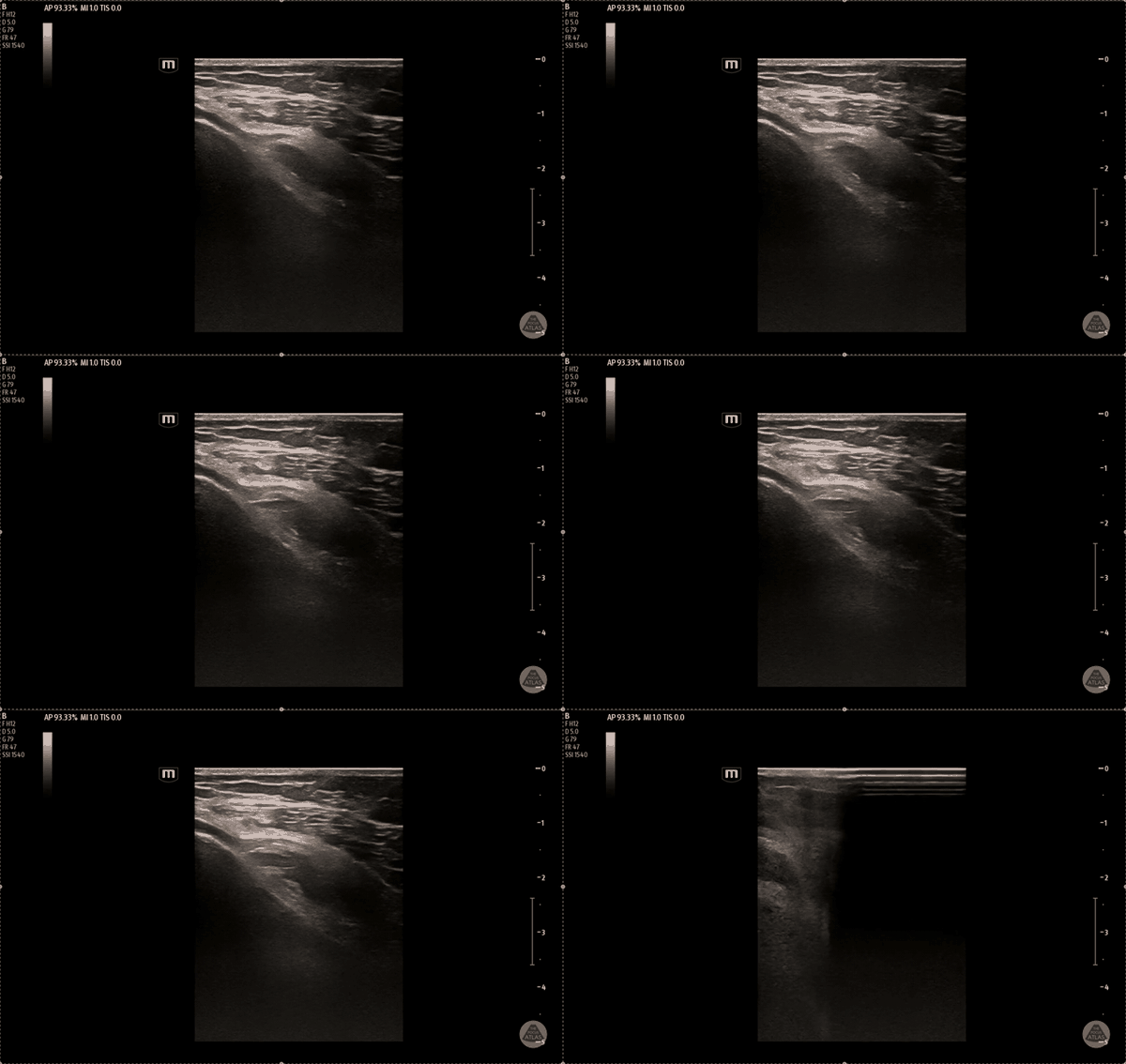
Indications
- Acute or chronic knee pain
Contraindications
- Infection overlying injection site
- Allergy to local anesthetic
- Avoid the inferolateral injection site due to proximity to the common peroneal nerve and risk of foot drop
Equipment
- 15cc of local anesthetic of choice
- Approximately 5cc per nerve
- 20-22G, 1.5-3.5in nerve block needle
- Cleansing solution
- Ultrasound with high-frequency linear transducer
- Ultrasound transducer sterile cover
Preparation
Position
- Position the patient supine
- Slightly flex the knee by placing a pillow in the popliteal fossa
Ultrasound
Landmarks
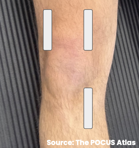
- Place the probe with the marker oriented cephalad
- Use a sagittal or slightly oblique plane
- Scan along:
- Medial distal femur
- Lateral distal femur
- Medial proximal tibia
- At each site, identify the long-bone metaphysis at the point of initial bony upsloping
- Look for adjacent genicular arteries when visible
- Identify the three injection targets:
- Superolateral genicular nerve distribution
- Superomedial genicular nerve distribution
- Inferomedial genicular nerve distribution
- Avoid the inferolateral site to prevent common peroneal involvement
Technique
- Use in-plane needle visualization
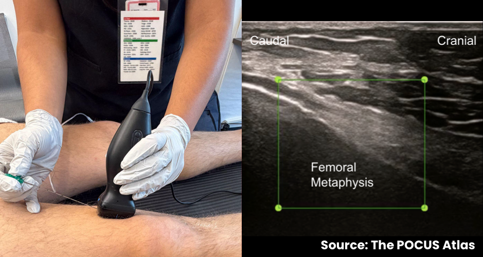
- For the superior nerves, advance the needle superior-to-inferior
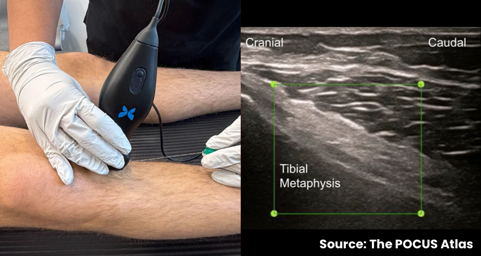
- For the inferior nerve, advance the needle inferior-to-superior
- Deposit approximately 5cc of local anesthetic against the periosteum at each site
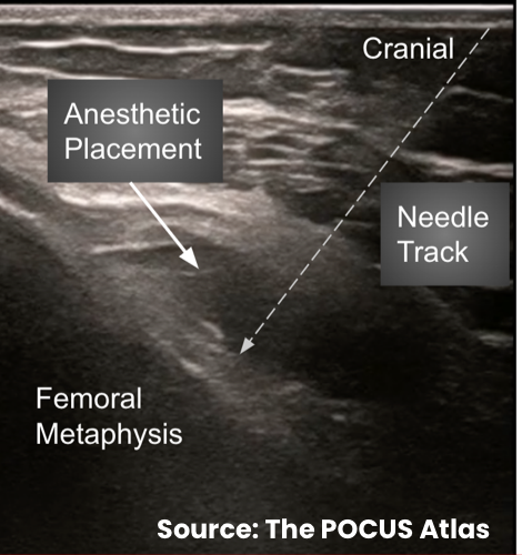
- Visualize anesthetic lifting the overlying tendons and musculature
- Repeat for all three target sites for complete joint coverage
Examples