Indications
- Knee surgery
- Medial knee pain
- Laceration, abscess, or foreign body removal of the medial lower leg or ankle
- Neuropathic pain in the saphenous nerve distribution
- Ankle fracture or reduction involving the medial malleolus
Contraindications
- Infection overlying injection site
- Allergy to local anesthetic
- Concern for compartment syndrome, especially in the setting of a tibial fracture
Equipment
- 10cc of local anesthetic of choice
- Calculate the maximum safe dose before performing the block to reduce the risk of local anesthetic systemic toxicity
- 20-22G, 5-10cm nerve block or short-bevel needle
- Cleansing solution
- Ultrasound with high-frequency linear transducer
- Ultrasound transducer sterile cover
Preparation
Position
- Document a neurovascular examination before performing the block
- Position the patient supine with the target leg abducted and externally rotated
Ultrasound
Landmarks
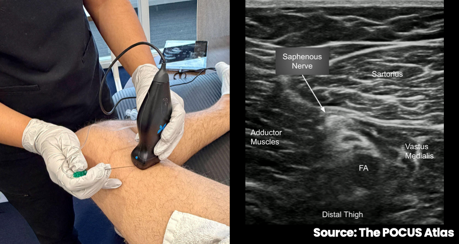
- Place the transducer transversely on the medial thigh over the adductor canal, approximately 5-7cm proximal to the popliteal crease
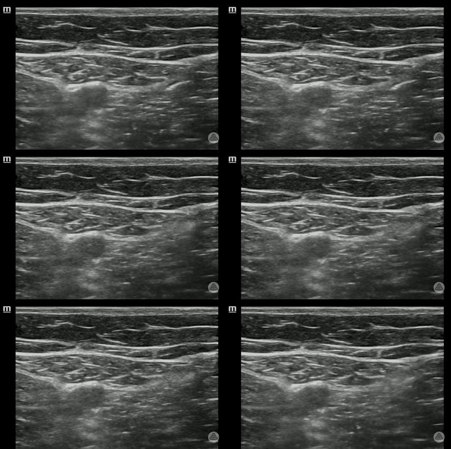
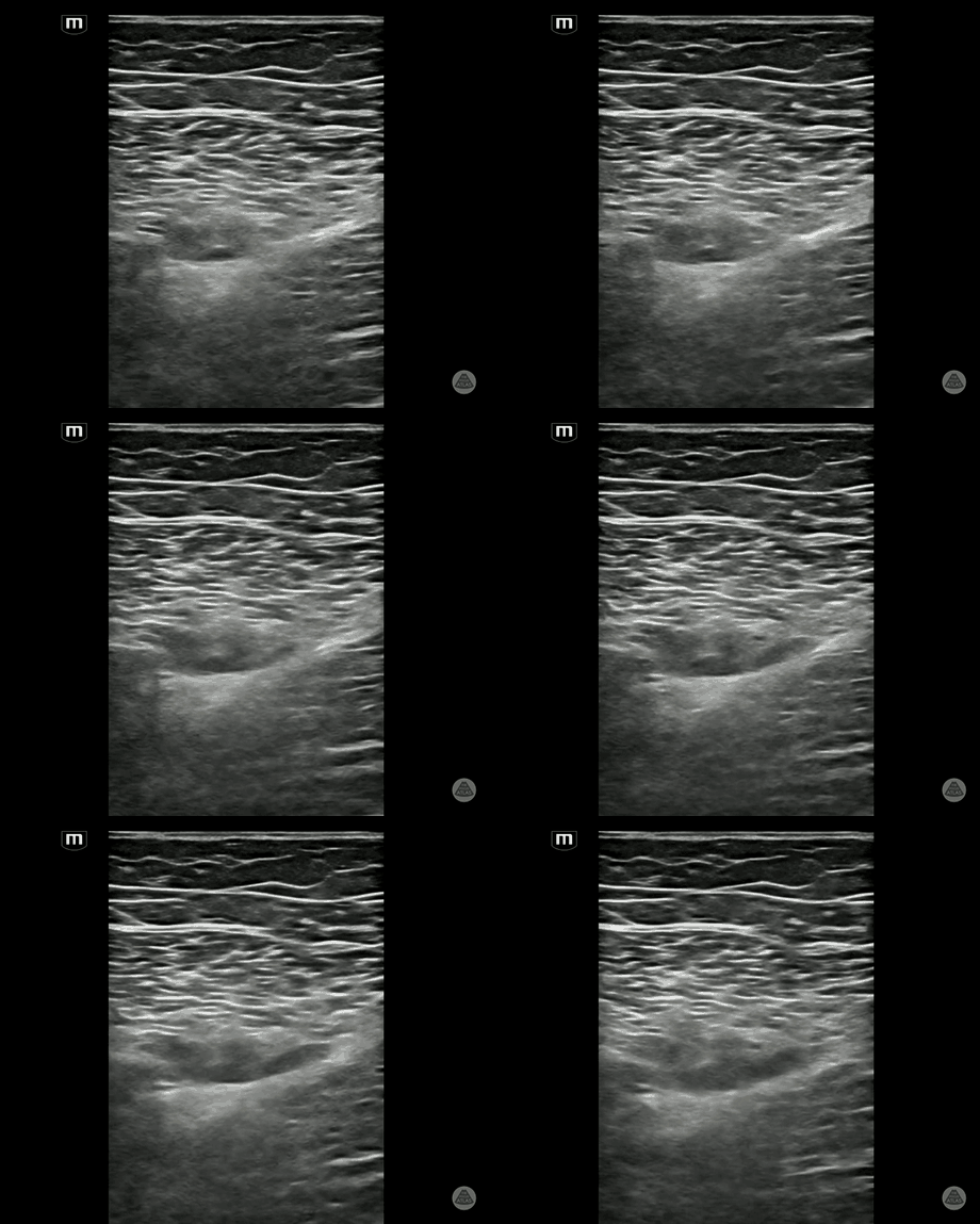
- Identify the sartorius muscle and the femoral artery
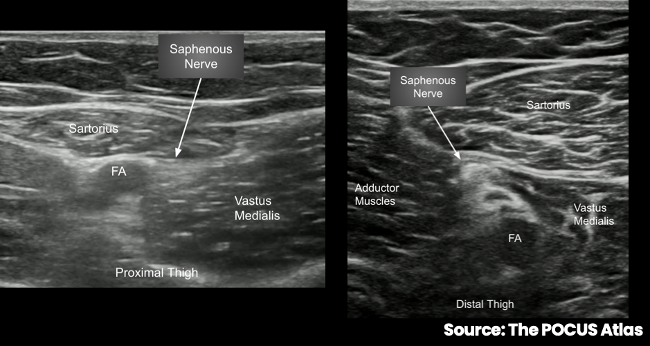
- Identify the saphenous nerve deep to the sartorius muscle and anterolateral to the femoral artery
Technique
- Advance the needle in-plane from medial to lateral
- Position the needle tip in the plane around the saphenous nerve
- Deposit approximately 10cc of local anesthetic and visualize spread around the nerve
- The block is primarily sensory, but mild vastus medialis weakness can occasionally occur
- Combine with a popliteal or distal sciatic nerve block when complete lower-extremity anesthesia is required
Examples